Intracellular Immunofluorescence Labeling Kit (INF-1)
Click to enlarge |
|
|
Intracellular Immunofluorescence Cell Staining (Labeling) Kit
Highlights
Suitable for 40 microscope slides
Kit Contents
Immunofluorescence Labelling of Cells
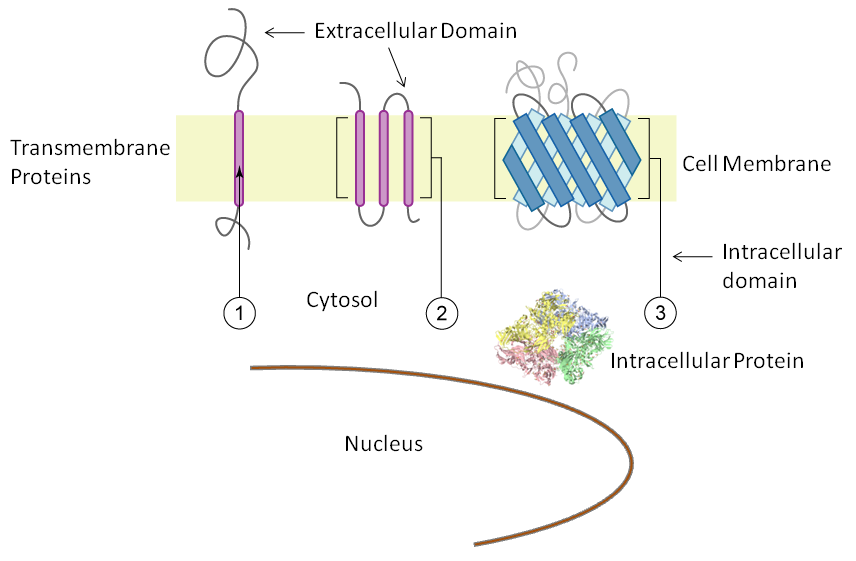
The Fivephoton Biochemicals intracellular immunofluorescence staining kit enables simultaneous labelling of multiple intracellular epitopes. The immunofluorescence kit uses bark saponin for membrane permeabilization, a reagent that permeates the outer cellular membrane as well as organeller membranes, and allows antibody to reach all cellular regions. Saponin permeation faciliates high resoluion imaging since membraneous structures remain intact. To exclusively label intracellular epitopes, use primary antibodies that recognize intracellular domains.
Immunofluorescence staining with membrane permeabilization is employed to visualize intracellular structures and organelles such as nuclei, actin filaments, Golgi, and endoplasmic reticulum. Intracellular cytosolic and organellar protein epitopes as well as intracellular domains of transmembrane proteins are labeled with this kit since antibodies have access to all intracellular regions.
(An important experimental distinction is provided in the Fivephoton Biochemicals Extracellular Immunofluorescence Kit (Part No. EIF-1) which enables exclusive extracellular labeling. The application in the Extracellular Immunofluorescence Labeling Kit does not include a membrane permeabilization reagent and therefore preserves membrane integrity).
Immunofluorescence Staining Protocol, Synopsis
Cells, grown on glass cover slips or slides, are first fixed in paraformaldehyde or methanol. The reaction is quenched with the provided "Quenching Buffer." Cells are then permeabilized with the provided "Permeabilization Buffer," and then incubated with antibodies using the included "Intracellular Detection Buffer." For multiple-labeling, up to three different antibodies generated in different hosts are added simultaneously in the primary antibody labeling step. After washes of unbound antibody, cells are incubated in the dark with fluorophore conjugated secondary antibodies. For multiple labeling, three matched pairs of secondary antibodies with different fluorescence tags are applied to the cells. Subsequent washes remove unbound antibody. Cover slips or slides are then mounted with an anti-fade solution. The anti-fade solution is allowed to harden and the cover slips or slides are ready for fluorescent microscopy.
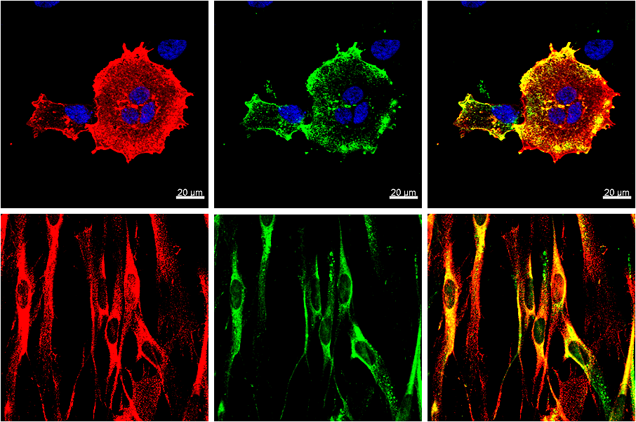
Multiple labeling immunofluorescence images developed with the Intracellular Immunofluorescence Kit (INF-1). Red: A cation channel. Green: An intracellular binding protein involved in membrane trafficking. Blue: DAPI staining of the nucleus.
Figure legends for "Images and Data" Tab
Representative microscopy images derived by the FIVEphoton Intracellular Immunofluorescence Labeling Kit are displayed in the icon and "Images and Data" tab. Microscopy images in the "Images and Data" tab are as follows: 1) An ABC transporter expressed by transfection in HEK cells: 2) Example of multiple-labeling immunofluorescence of an amyloid protein and an endoplasmic reticulum marker expressed by transfection in COS-7 cells.
Cellular Regions and Membrane Topology
Representative References on Immunofluorescence Labeling
Safety: Contains Irritants. Avoid ingestion, eye and skin contact.
Storage: Shipped at ambient temperature. Store at -20oC. Solutions can be thawed and frozen multiple times.
Shipping Options: Fedex 2-3 day domestic delivery or overnight delivery. Contact salessupport@FIVEphoton.com for other delivery options, including international delivery. |
 Products
 Manuals


